Thursday, November 15, 2007

Multicystic Teratoma

A 27 y/o G0P0 presented with oligomenorrhea that started a year ago.
She was given combination pills and pelvic US which revealed 10 cm complex R adnexal mass.
No abdominal pain/fullness, weight loss or anorexia.
Menarche at age 13 with no hx of abnormal Pap or chlamydia/trich.
Obesity was noted.
Disorganized endocervical stroma with breakdown per endometrial bx.
Elevated LDH - 192.


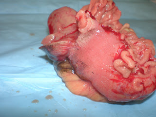
R oophorectomy specimen. Serous fluid when opened.

So when encountering teratoma,
1) if mature, then no further treatment needed.
2) if immature component seen in histology, treatment more aggressive. More lymph node dissection and adjuvant therapy.

In immature teratoma, malignancy mostly arise from neural component.


This specimen shows multiple dermoid cysts each spitting out sebum with hair when opened.


There was also a tooth(that dark spot) where I palpated. Either tooth or just calcification.

Frozen section showed sebaceous glands, smooth muscle, ovarian stroma, pancreas, colon, brain, hobnailing area that looked choroid plexus(?) and small lymphoid cells but had clear vacuole in the nucleus. Pleomorphic and less differentiated, diagnosis was mature teratoma with focal area suggestive of immature element.


Ovarian stroma


Sebaceous glands with hair shaft and smooth muscle around.


fibroadenomatous.


tubal


intestinal


pancreatic acini


neural

so far these looked well differentiated until we reached a focus around neural tissue.

Tuesday, November 13, 2007

Anemia classification by MCV

1) Microcytic, hypochromic (MCV < style="font-weight: bold;">iron deficiency
Thalassemias
Lead poisoning, sideroblastic anemias

2)Macrocytic (MCV > 100)
Megaloblastic - vitamin B12/folate deficiency
DNA synthesis inhibitor (sulfa drug, AZT)
Marked reticulocytosis(reticulocytes bigger than RBC)

3)Normocytic, normochromic
Acute hemorrhage
Enzyme defects (G6PD deficiency, PK deficiency)
RBC membrane defects (spherocytosis)
Bone marrow disorder (aplastic anemia, leukemia)
sickle cell disease
autoimmune hemolytic anemia
anemia of chronic disease: decreased total iron binding capacity, increased ferritin, increased storage of iron in marrow macrophage

anemia note

Notes on Hematology

Friday, November 9, 2007

Uterine Polyp: Hysterectomy specimen


1) Hysterectomy specimen before inking for margins.
Can see:
- cervix, cervical canal, corpus, fundus, fallopian tubes, serosal leiomyomata


3) section before being processed. The polyp is superficial. Endometrial lining (which is replaced by polyp in this image)is supposed to be very thin (few milimeters?), the pt was past 60.


2) Can see the jelly-like polyp in the midst of fibroid field. This polyp looked pretty benign bit hyperplastic in sections but in cytology cells looked very malignant. Also the previous history of endometrial papillary serous carcinoma that was resected was recalled. Thus the diagnosis is same (even without histology to prove it), malignant per cytology.

Friday, November 2, 2007

Petechial hemorrhage on sigmoid colon


These hemorrhagic petechiae in the background of mucosal erythema may appear melanosis coli.


Sigmoidal polyp is present, possibly tubular adenoma.

GIST


This serosal lesion on small intestine is GIST. GISTs are different from leiomyoma in gross appearance. While leiomyoma have white pale fibrosed lesion, GISTs tend to have burgundy color.


The spherical GIST was split half, mucosa exposed underneath. Grossly the lesion didn't seem to involve mucosa.


Touch prep shows abundant spindle cells. Leiomyoma will barely have cells on touch prep, unlike GIST.


Frozen section image.