Thursday, July 5, 2007

Diabetes

Primary

- Type I (insulin dependent, IDDM)

- Type II (non-insulin dependent, NIDDM)

non-obese NIDDM

obese NIDDM

- Maturity-onset diabetes of the young (MODY) - genetic defect of Beta cells


Secondary

- chronic pancreatitis

- post pancreatectomy

- hormonal tumors (pheochromocytoma, pituitary tumor)

- drugs (corticosteroids)

- hemochromatosis

- genetic disorder


Glucose + GLUT-2 signals Beta cells to produce insulin


Insulin signals target cells to produce GLUT-4 for glucose transport


Type I IDDM

- HLA-D gene (DP, DQ, DR) - class II MHC renders Beta cells immunogenic


Type II NIDDM

- derangement in Beta cell secretion of insulins

- decreased response of peripheral tissues to respond to insulin

- also leads to irreversible Beta cell injury

- obesity, insulin insensitivity

- gestational diabetes (obesity, pregnancy)

- amylin - normally produced with Beta cells copackaged with insulin

- in NIDDM, accumulate in sinusoidal space outside the Beta cell

=> eventually become like amyloid


amyloid - insoluble fibrous protein aggregation

- physically disrupt tissue architecture


Complications of diabetes (seen in both type I and II)

- microangiopathy, retinopathy

- nephropathy, etc.


Nonenzymatic glycosylation

- degree of enzymatic glycolytation at blood glucose

Reversible vs. Irreversible glycolylation

irreversible advanced glycosylation end products (AGE)

early glycosylation products rearrange intead of dissociating

AGE crosslinking with LDL in blood vessel or albumin in renal glomeruli

-> decrease protein removal, increase protein deposit

AGE binding to receptors of blood cells

-> procoagulation

Tumors of Liver

Harmatoma - benign tumor composed of tissue elements found at site but disorganized

Benign
- hepatic adenoma
- histologically no portal structure but with normal liver apperance
- ~ 20 cm well circumscribed nodules, predisposed to increased estrogen
- bile duct adenoma
- small white nodules beneath liver capsule
- abnormal "bile duct" in a collagenous stroma
- hemangioma
- beneath capsule as a dark/black lesion 2~3 cm
- histologically abnormal "vascular channels" in a collagenous stroma

Malignancy
- metastatic tumor (lung, colon, breast, stomach), malignant lymphomas, leukemias
=> accumulation of tumor deposits can lead to compression of intrahepatic bile duct and lead to obstructive jaundice
- primary hepatocellular carcinoma (hepatoma)
risks - cirrhosis, HBV, mycotoxins
Serum Alpha-fetoproteins may be raised
- cholangiocarcinoma
- due to chronic inflammatory disease of intra-hepatic biliary tree
- angiosarcoma
- multifocal hemorrhagic nodules

Pancreatitis and Islet Cell tumors


Pancreatitis
1) Periductal Obstruction -> necrosis
2) Perilobar Necrosis due to ischemia
3) panlobular necrosis with hemorrhage from vessels

Interstitial inflammation, edema, proteolysis, fat necrosis, hemorrhage

PANCREAS ARISE FROM 2 DUODENAL FOLDS AS DORSAL AND VENTRAL PANCREAS THAT FUSE TO BECOME ONE

Activation of trypsinogen an important triggering event in pancreatitis

ACUTE PANCREATITIS
- SYSTEMIC ORGAN FAILURE
SHOCK
ARDS
ACUTE RENAL FAILURE
- PANCREATIC ABSCESS
- PANCREATIC PSEUDOCYST
- DUODENAL OBSTRUCTION

Pseudocyst - unilocular; multiloculation suggests a neoplastic cyst
- virtually all arise after acute/chronic pancreatitis
Cyst
- lined by flattened cuboidal epithelium with clear glycogen rich cytoplasm

Pancreatic Carcinoma
- virtually all cancers begin in the ductal epithelium

60% head of pancreas, earlier detection
15-20% body
5% tail

--------------------------------------------------------

ENDOCRINE PANCREAS
ALPHA - GLUCAGON
BETA - INSULIN
DELTA - SOMATOSTATIN
PP - PANCREATIC POLYPEPTIDE
small dark granules present in islets as well as in exocrine pancreas

--------------------------------------------------------

CHRONIC PANCREATITIS, POSSIBLE CAUSES
1) DUCTAL OBSTRUCTION BY CONCRETIONS
2) SECRETED PROTEINS - LITHOSTATHINE
3) OXIDATIVE STRESS
EtOH induced
- free radical in acinar cells
- abnormal protein secretion
- acinar cell necrosis, inflammation, fibrosis
- interstitital fibrosis
ACUTE PANCREATITIS -> NECROSIS -> FIBROSIS
also, abnormal intrapancreatic lipid metabolites

--------------------------------------------------------

Islet cell tumors
1) hyperinsulinism (insulinoma) - Beta cell tumor
2) hypergastrinemia / Zollinger-Ellison syndrome
3) multiple endocrine neoplasia

MEN I (for 1 and 2)
1) hyperinsulinism
- blood glucose < 50 mg/dL hypoglycemic attack
- CNS manifestation
- resolve with increased glucose
- high insulin glucose ratio
2) gastrinomas
- hypersecretion of gastrin with severe peptic ulceration
ulcer (90~95%) duodenal : gastric = 6 : 1
- diarrhea
- in duodenum, peripancreatic soft tissues or pancreas
Treatment - control of gastric acid secretion by histamine (H2) receptor blockers, resection

3) other islet cell tumors
alpha cell tumor - glucagonomas
delta cell tumor - somatostatinoma
VIPomas - VIP
pancreatic carcinoid tumors - serotonin

Liver, general dz characteristics

LFT enzymes
Liver Necrosis
Liver Disease Characteristics
Vascular Liver Dz
Acute vs Chronic Hepatitis
Bacterial Infection Route
Patterns of Chronic Hepatitis
Metabolic Liver Disease - hemochromatosis, Wilson's dz
Cirrhosis
Biliary Cirrhosis
=======================================
main rate limiting step in bilirubin metabolism is excretion by the canaliculi rather than conjugation

- alkaline phosphatase - located on the cell membrane of biliary canaliculi
- transaminase - located in the hepatocyte cytoplasm, ALT more specific for liver than AST
- conjugated bilirubin - secreted by liver cells associated with biliary obstruction or liver cell destruction
- albumen - reflects synthetic property of liver, low level => long standing disease of liver
- caeruloplasmin
- transferrin


space of Disse
between hepatocyte surface and endothelial lining of sinusoid (larger than capillaries)


Liver Necrosis
- councilman bodies - dead hepatocytes form eosinophilic shrunken structure
- spotty necrosis - patchy
- zonal necrosis - necrosis confined to zone
- piecemeal necrosis - scattered pattern immediately next to the portal-tract connective tissue
- bridging necrosis - extensive necrosis that bridge different veins/tracts
General Liver Disease Characteristics
- fatty change
- cholestasis
- extrahepatic
- intrahepatic
- liver necrosis
- liver fibrosis - cells of Ito in space of Disse
- liver storage disease
- hemochromatosis, hemosiderosis, Wilson's disease
- glycogenesis (glycogen storage dz)
Vascular liver disease
- true infarct - rare, from trauma, arterial embolization, bacterial endocarditis, eclampsia, polyarteritis nodosa(Kussmaul disease)
- R sided heart failure -> passive venous congestion of liver -> nutmeg appearance (chronic passive venous congestion)
- portal HTN
- increase in portal venous pressure
=> splenomegaly, ascites
- new channels may open up betweeen portal and systemic venous circulation
- variceal bleeding
- caput medusa
- hemorrhoids
- classified: pre-sinusoidal, sinusoidal or post-sinusoidal
EXAMPLES: PRE-SINUSOIDAL - PORTAL VEIN THROMBOSIS
SINUSOIDAL - HEPATIC FIBROSIS, CIRRHOSIS
POST SINUSOIDAL - HEPATIC VEIN THROMBISIS, CENTRAL VEIN THROMBOSIS
Clinical Picture: Budd-Chiari Syndrome (post sinusoidal)
TRIAD: 1) ABD PAIN
2) ASCITES
3) HEPATOMEGALY
--------------------------------------------------------
ACUTE HEPATITIS
- INCREASED BILIRUBIN
- INCREASED ALT/AST - LIVER CELL NECROSIS
- ALBUMEN ~ NORMAL
- DECREASED COAGULATION DUE TO DECREASE IN COAGULATION FACTOR PRODUCTION
Hepatrophic Virus
A, E = fecal, oral
B, C, D = parenteral, histologically ground glass apperance of hepatocytes (accumulation of antibodies)
Route of Bacterial Infection
- ascending from biliary tract
- ascending in the portal vessels from a focus of sepsis in abdomen
- systemic septicemia
3 histological Patterns of Chronic Hepatitis
- chronic active hepatitis
- continued necrosis of hepatocytes
- development of cirrhosis
- necrosis extends from one portal area to another, portal tract to parenchyma
- chronic persistent hepatitis
- confined to portal tract
- not associated with progressive fibrosis or cirrhosis
- chronic lobular hepatitis
- portal tract inflammation(no piecemeal necrosis), spotty parenchymal inflammation
Metabolic Liver disease
Iron - hemochromatosis
primary - excessive absorption of iron(accumulates as hemosiderin) from gut
- chromosome 6, HLA locus
- cells look rusty brown due to hemosiderin in cells
- great increase transferrin in blood, increase of iron, ferritin
secondary - also called hemosiderosis
- due to other disease (alcoholism) or repeated blood transfusion
Copper - Wilson's disease
- decrease in ceruloplasmin (Cu binding) in blood
- liver fails to excrete
- Cu-ceruloplasmin complex
- overspills into blood, deposited in brain, cornea
Cirrhosis hx
- long destruction of liver cell
- chronic inflammation stimulating fibrosis
- regeneration of hepatocytes to form nodules
Biliary Cirrhosis
- Secondary - obstructed extrahepatic duct
- Primary - Autoimmune, Slow destruction of bile canaliculi
- sclerosing cholangitis
- associated with inflammatory bowel disease
- both intra/extra hepatic duct
- medium sized ducts, ducts in portal tract => concentric fibrosis and inflammation, small ducts => replaced by collagen, fibrous stricture with segmental dilatation

Breast Pathology NOT COMPLETE

Wednesday, July 4, 2007

Innate vs. Adaptive Immunity


INNATE - neutrophil, macrophage, dendritic cells, complement
ADAPTIVE - T, B-cells, circulating antibodies

T-cell differentiation (figure)

T-cell activation
MHC - signal 1
recognized by TCR
B7-CD28, IL-2 - signal 2
costimulatory molecule
activation requires 2 signals


Lymphocytes

T cell (60~70%)
- CD4+ help B cells make antibody and activate macrophage
- CD8+ kill virus infected cells directly
- allergy (type IV sensitivity)
- slow organ rejection
B cell(10~20%); found in superficial cortex of lymph nodes, white plulp in spleen
- make antibody, form plasma cells
- IgG antibodies to oppose bacteria, viruses
- allergy (type I hypersensitivity); IgE
- antibodies reject organ fast

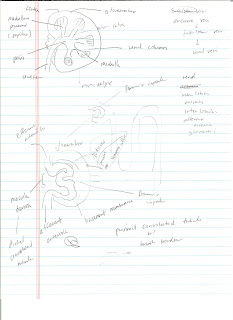
Kidney Anatomy

Herpes Virus

Herpes Virus
- double stranded DNA ~ 70 proteins
- types
a. neutropic alpha-group viruses
HSV1, HSV2, VZV
b. lymphotropic beta-group viruses
CMV, HHV6, HHV7
c. gamma group viruses
EBV, HHV8 (Kaposi's Sarcoma)

HSV1, HSV2
- cause vesicular lesions of the epidermis
- infect neurons
- primary HSV infection in immunocompetent resolve in few weeks (VIRUS LATENT IN NERVE CELLS)

HSV morphology
- formation of large, pink to purple (Cowdry type A) intranuclear inclusions that contain intact and disrupted virions and push darkly stained host cell chromatins to the edge of nucleus
inclusion-bearing multinucleated syncitia

Manifest
- cold sore, fever blister
- gingivostomatitis
- genital herpes
- corneal lesions (herpes epithelial keratitis, herpes stromal keratitis)